Can an X-Ray Show Cancer in the Bones?
2/16/2026
If you or someone you care about has persistent bone pain or an abnormal imaging result, you may be wondering: Can an X-ray show cancer in the bones? The short answer is yes — but with limitations. X-rays can sometimes detect bone cancer or metastatic cancer that has spread to the bones, but they are usually not the only test doctors rely on for diagnosis.
In this guide, we’ll explain how X-rays detect bone cancer, what signs radiologists look for, when additional imaging is required, and what to expect during evaluation.
How X-Rays Work
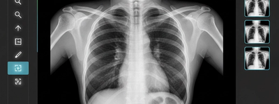
An X-ray is a form of imaging that uses low-dose ionizing radiation to create pictures of structures inside the body. Dense materials like bone absorb more radiation and appear white on the image, while softer tissues appear darker.
Because bones are dense and clearly visible on X-rays, abnormalities in bone structure can often be detected early.
Can an X-Ray Detect Bone Cancer?
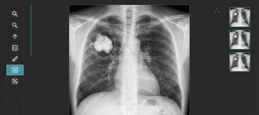
Yes, X-rays can sometimes reveal signs of bone cancer. However, they do not directly “see” cancer cells. Instead, they show structural changes in the bone that may suggest a tumor.
What an X-Ray Might Show
If bone cancer is present, an X-ray may reveal:
- Areas of bone destruction (osteolytic lesions)
- Abnormal bone growth (osteoblastic lesions)
- Irregular bone shape or structure
- Bone thinning or weakening
- Unexplained fractures
- Soft tissue swelling near the bone
These changes raise suspicion, but they do not confirm cancer. Further testing is almost always required.
Types of Bone Cancer
There are two main categories doctors consider:
1. Primary Bone Cancer
This begins in the bone itself. Examples include:
- Osteosarcoma
- Ewing sarcoma
- Chondrosarcoma
Primary bone cancers are rare.
2. Secondary (Metastatic) Bone Cancer
This is much more common. It occurs when cancer from another part of the body spreads to the bones. Common primary cancers that spread to bone include:
- Breast cancer
- Prostate cancer
- Lung cancer
- Kidney cancer
- Thyroid cancer
X-rays are often one of the first imaging tests used when metastasis is suspected.
When Is an X-Ray Ordered?
A doctor may recommend an X-ray if you have:
- Persistent bone pain
- Swelling near a bone
- A lump or mass
- Unexplained fractures
- Limited movement in a limb
- A history of cancer with new bone pain
X-rays are typically the first step because they are quick, accessible, and cost-effective.
Limitations of X-Rays in Detecting Bone Cancer
Although X-rays can show suspicious bone changes, they have important limitations:
1. Early Cancer May Not Be Visible
Small tumors or early-stage bone cancer may not produce noticeable structural changes.
2. Soft Tissue Detail Is Limited
X-rays are excellent for bone but poor at evaluating soft tissues and bone marrow.
3. Cannot Confirm Diagnosis
An X-ray cannot determine whether a lesion is cancerous, benign, infectious, or caused by another condition.
Because of these limitations, abnormal findings are usually followed by additional tests.
What Happens If an X-Ray Shows Something Abnormal?
If your X-ray reveals suspicious findings, your doctor may recommend:
- MRI (Magnetic Resonance Imaging): Provides detailed images of bone marrow and surrounding soft tissues.
- CT scan: Offers cross-sectional views for better structural detail.
- Bone scan: Detects areas of increased bone activity.
- PET scan: Identifies areas of high metabolic activity typical of cancer.
- Biopsy: A small sample of tissue is removed and analyzed under a microscope. This is the only way to confirm cancer.
In many cases, what appears suspicious on X-ray turns out to be a benign bone cyst, infection, or non-cancerous growth.
What Does Bone Cancer Look Like on an X-Ray?
Radiologists are trained to look for specific patterns. Some common appearances include:
- “Moth-eaten” bone destruction
- Periosteal reaction (new bone forming around a lesion)
- Sunburst pattern (spiculated bone growth)
- Codman triangle (elevation of periosteum)
- Mixed lytic and sclerotic areas
These patterns do not automatically mean cancer, but they guide further investigation.
Symptoms of Bone Cancer
While imaging is crucial, symptoms often prompt testing. Warning signs may include:
- Persistent, worsening bone pain (especially at night)
- Swelling or tenderness
- A visible lump
- Fatigue
- Unexplained weight loss
- Frequent fractures
However, many of these symptoms are caused by non-cancerous conditions.
Is an X-Ray Enough to Diagnose Bone Cancer?
No. An X-ray alone is not sufficient to diagnose bone cancer. It serves as an initial screening tool.
Diagnosis typically involves:
- Imaging (X-ray first)
- Advanced imaging (MRI or CT)
- Laboratory tests
- Biopsy confirmation
Only a biopsy can definitively confirm cancer.
What to Expect During the X-Ray
If your doctor orders an X-ray to evaluate possible bone cancer, the process is simple:
Before the Exam
- Remove jewelry or metal objects.
- Inform the technician if you are pregnant.
During the Exam
- The technician positions the affected body part.
- You remain still for a few seconds.
- The image is captured in seconds.
The entire appointment usually takes less than 20 minutes.
After the Exam
- No recovery time is needed.
- A radiologist reviews the image.
- Your doctor discusses the results and next steps.
Radiation Safety
X-rays use a small amount of radiation, but exposure levels are medically regulated and considered safe for diagnostic use. The benefits of detecting serious conditions generally outweigh the minimal risks.
However, unnecessary repeated imaging should be avoided unless medically justified.
Final Thoughts
So, can an X-ray show cancer in the bones? Yes — it can reveal structural changes that raise suspicion. However, it cannot definitively diagnose cancer on its own. X-rays are typically the first step in a larger diagnostic process that may include MRI, CT scans, bone scans, and biopsy.
If you’re experiencing persistent bone pain or have concerns about imaging results, the most important step is to discuss them with your healthcare provider. Early evaluation and proper follow-up testing are key to accurate diagnosis and effective treatment.
Frequently Asked Questions
Not always. Early bone cancer may not cause visible changes on X-ray. MRI is more sensitive for early detection.
No. If symptoms persist despite a normal X-ray, further imaging may still be required.
Primary bone cancer is rare. Metastatic bone cancer (spread from another cancer) is more common.
Certain conditions like arthritis or infections can sometimes mimic suspicious changes, which is why additional tests are needed.
If imaging strongly suggests cancer, a biopsy is usually required to confirm the diagnosis.
In urgent cases, results may be reviewed the same day. Otherwise, it typically takes 24–48 hours.
Not necessarily. Many abnormal X-ray findings turn out to be benign conditions.