X-Ray of the Ankle: How It Identifies Sprains, Fractures, and Joint Issues
2/11/2026
An ankle injury can happen in a split second—stepping off a curb awkwardly, twisting during sports, or slipping on uneven ground. When pain, swelling, or trouble walking follow, a doctor often orders an ankle X-ray to understand what’s going on inside. Ankle X-rays are among the most common imaging tests in orthopedics. They play a crucial role in diagnosing fractures, evaluating the joint structure, and helping decide whether more testing is needed.
In this article, you’ll learn how ankle X-rays work, what they can and can’t show, and why they’re often the first step in diagnosing ankle injuries. The goal is to make the topic easy to follow, even if you’re not a medical expert.
What an Ankle X-Ray Looks At
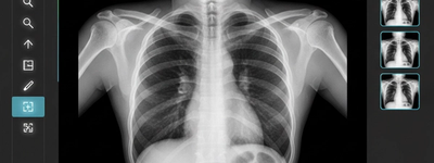
An X-ray is a simple imaging test. It uses a small amount of radiation to take pictures of your ankle bones and joints. These images show the shape, position, and structure of your bones in different views.
Bones and Major Joint Elements
When you get an ankle X-ray, the machine captures images of the three main bones:
- Tibia – the larger shinbone
- Fibula – the smaller bone next to the tibia
- Talus – the small bone that sits between the lower leg and the heel bones
In a healthy ankle X-ray, these bones appear smooth, well-aligned, and uninterrupted. The spaces between them—the joint spaces—are even and clear, which usually reflects good cartilage health.
How X-Rays Detect Fractures
One of the main reasons doctors order an ankle X-ray is to check for a fracture—a break in one or more bones. On an X-ray:
- Fracture lines appear as dark lines or gaps through the bone.
- Bone displacement shows bones that are shifted or not aligned normally.
- Bone fragments may be visible if the break is complex.
Fractures can be simple (a clean break) or complex (involving multiple breaks or displacement). Some fractures are easy to see on a routine X-ray. Others, like very small cracks or injuries hidden in tricky areas, may need additional views or follow-up imaging to be detected.
Sprains: What X-Rays Can Do and Can’t Do
A sprain happens when ligaments—the strong bands of tissue holding bones together—are stretched or torn. These soft tissues do not show up on a standard X-ray. That means an X-ray cannot directly show a sprain or a torn ligament.
So why do doctors still order X-rays for sprains?
To Rule Out Fractures
Sprains and fractures can feel very similar. Both can cause pain, swelling, and difficulty walking. X-rays help doctors rule out a broken bone, which requires different treatment than a ligament injury.
Indirect Clues
Sometimes an X-ray can hint at ligament problems. For example:
- Avulsion fractures, where a piece of bone is pulled off by a ligament, show up on X-rays and may suggest a severe ligament injury.
- Joint widening or subtle changes in bone alignment might suggest associated soft-tissue damage.
Limitations of X-Rays for Sprains
Because ligaments and other soft tissues aren’t directly shown, many sprains—especially mild ones—appear normal on X-rays. If pain and dysfunction continue after a normal X-ray, a doctor might recommend an MRI or an ultrasound to look more closely at ligaments, tendons, and cartilage.
Joint and Degenerative Changes
Ankle X-rays also reveal changes beyond acute trauma:
Arthritis and Degeneration
If someone has long-term ankle pain—especially with age or past injuries—X-rays may show signs of arthritis. These can include:
- Narrowed joint spaces
- Bone spurs
- Irregular bone surfaces
These findings help doctors understand chronic conditions and plan appropriate treatments.
Dislocations or Misalignments
Sometimes bones are pushed out of place within the joint. An X-ray clearly shows this misalignment, which often requires urgent medical care.
Different X-Ray Views and Why They Matter
A standard ankle X-ray isn’t just one picture. Technicians take several views:
- Anteroposterior (AP) – from the front
- Lateral – from the side
- Mortise view – taken at a specific angle to show the entire joint without overlap
The mortise view is especially useful. It shows the entire joint space and helps doctors spot issues that might be missed on straight-on or side views. It’s often used when injuries are subtle or complex.
When Are Ankle X-Rays Recommended?
Doctors don’t automatically X-ray every ankle injury. They use clinical guidelines, like the Ottawa ankle rules, to decide when it’s necessary. These rules help determine who really needs an X-ray, minimizing unnecessary radiation and delays in care.
Some common justifications for imaging include:
- Pain in key bone areas after trauma
- Inability to bear weight
- Significant swelling or deformity
- Strong suspicion of a fracture
What Happens After the X-Ray?
Your doctor interprets the images and explains what they mean. If the X-ray shows a fracture, the treatment plan may include casting, splinting, or even surgery, depending on severity. If no bone injury is seen but symptoms are significant, your doctor might recommend further imaging or physical therapy for ligament damage.
Remember: a normal X-ray doesn’t always mean you’re fine. Persistent pain, instability, or swelling may still require follow-up imaging.
Conclusion: Why Ankle X-Rays Matter
An ankle X-ray is a fast, accessible first step in diagnosing many types of injuries. It helps doctors:
- Identify fractures and bone breaks
- Assess joint alignment and spaces
- Spot signs of arthritis or chronic changes
- Rule out serious bone damage when sprains are suspected
However, it has limits. X-rays do not show ligaments, cartilage, or tendon injuries directly, so sometimes additional imaging is needed to fully understand soft-tissue damage.
If you’re ever unsure about your diagnosis or recovery path, it’s okay to ask your healthcare provider to explain your X-ray findings in plain language. Understanding what the images show helps you manage your injury with confidence.
Frequently Asked Questions
Technically, an X-ray identifies a sprain by exclusion. Because ligaments (which are stretched or torn in a sprain) are soft tissue, they don't show up on a standard X-ray. If the X-ray shows the bones are perfectly intact and aligned, the doctor will likely diagnose a sprain. If the X-ray shows a break in the malleolus (the "knobs" on the side of your ankle) or the talus, it is a fracture.
Sometimes a doctor will ask you to stand up while the X-ray is taken. This is called a Weight-Bearing view. It is crucial for identifying joint instability or "syndesmosis" injuries (high ankle sprains). By putting your body weight on the ankle, the doctor can see if the space between your bones widens under pressure, which indicates ligament damage that wouldn't be visible while you are sitting or lying down.
Yes. For chronic pain, an X-ray looks for signs of wear and tear. Key indicators include: Joint Space Narrowing: The cartilage has worn down, bringing the bones closer together. Osteophytes (Bone Spurs): Small bony growths that form as the body tries to stabilize a damaged joint. Sclerosis: Areas where the bone has become unusually dense or "scarred" due to constant friction.
To get a complete picture, the technician usually performs a "Three-View Series": AP (Anteroposterior): Your foot points straight up. Lateral: You turn on your side so the technician can see the ankle from the profile. Mortise View: You rotate your foot slightly inward (about 15 degrees). This view is the most important for looking at the "mortise"—the square-shaped hole where the leg bones meet the ankle bone.
It is a common misconception that a "clear" X-ray means everything is fine. A clear X-ray only means the bones are okay. You could still have: Grade III Sprains: A complete tear of the ligaments. Occult Fractures: Tiny "stress fractures" that are too small for a standard X-ray to pick up in the first few days. Cartilage Damage: Tears in the lining of the joint. In these cases, your doctor might recommend an MRI or CT scan for a more detailed look at the soft tissues.