Shoulder X-Ray: Anatomy, Procedure & What to Expect
2/11/2026
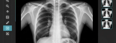
When you hear that you need a shoulder X-ray, you may feel nervous or unsure about what will happen. A shoulder X-ray is one of the most common imaging tests doctors use to look at the bones and joints of the shoulder. It’s fast, safe, and gives valuable information that helps diagnose injuries and other conditions. This guide will walk you through what a shoulder X-ray shows, why it’s done, how it’s performed, and what you can expect before, during, and after the procedure.
Understanding the Shoulder Anatomy
The shoulder is one of the most mobile joints in the body. It is built to let you lift your arm, reach overhead, and rotate your hand in many directions. Several key bones form this complex joint:
- Humerus — the upper arm bone with a rounded head that fits into the shoulder socket.
- Scapula (Shoulder Blade) — a flat bone that provides the socket (glenoid) for the humerus.
- Clavicle (Collarbone) — connects the shoulder to the sternum and helps stabilize movement.
- Acromion — a bony projection off the scapula.
These bones make up the glenohumeral joint, often referred to as the shoulder joint. Cartilage, muscles, and tendons around these bones help support and move the joint, though they don’t show up clearly on X-rays.
Why Doctors Order a Shoulder X-Ray
A shoulder X-ray is usually the first imaging test used to evaluate shoulder pain, injury, or functional problems. It helps doctors see the shape, position, and integrity of the bones in your shoulder joint.
Here are common reasons a healthcare provider might recommend this test:
- Pain or Discomfort — persistent pain that doesn’t resolve on its own.
- Injury or Trauma — after a fall, blow, or accident to check for fractures or dislocations.
- Limited Range of Motion — difficulty moving the shoulder in normal directions.
- Swelling or Deformity — visible changes in the shoulder’s appearance.
- Suspected Arthritis — to look for changes in bone structure from wear or inflammation.
X-rays can also reveal bone spurs, tumors, and signs of infection around the bones. While an X-ray can show bone problems clearly, it offers limited detail about soft tissues like muscles and tendons — for that, an MRI or ultrasound may be requested later.
Preparing for Your Shoulder X-Ray
Luckily, a shoulder X-ray requires little to no preparation. You do not need to fast or change your normal routine. However, your technologist will ask you to remove any clothing or jewelry that might interfere with the images.
Before the test, share important details with your provider, such as:
- Pregnancy — X-rays use a small amount of radiation, and they are generally avoided during pregnancy unless essential.
- Previous Imaging — if you have recent shoulder X-rays, bring those images along so doctors can compare them.
- Medical History — tell your provider about any metal implants, pins, or plates near the area to be X-rayed.
Wear comfortable, loose clothing to your appointment. You may be asked to change into a medical gown.
What Happens During the Shoulder X-Ray
A shoulder X-ray is quick, typically taking 10–15 minutes from start to finish, including positioning and image capture.
Here’s what you can expect:
- Positioning — You will sit, stand, or lie down based on the view the technologist needs.
- Multiple Views — The X-ray technician will take images from different angles, such as front (anteroposterior), side (lateral), and sometimes oblique views, to fully visualize the shoulder.
- Stillness — You’ll be asked to hold still and sometimes hold your breath briefly while the machine takes pictures.
You won’t feel pain from the X-ray itself. Some discomfort may arise if you have pain in the shoulder being imaged and have to hold certain positions.
After the X-Ray: Results and Next Steps
Once the images are taken, your technologist will check them to make sure they are clear. If not, a second set may be needed. A radiologist (a doctor trained in interpreting medical imaging) reviews the images and shares findings with the provider who ordered the test.
In many cases, results are available within minutes to a few days. Based on the findings, your doctor will discuss a treatment plan, which might include medication, physical therapy, further tests like MRI, or surgical options if needed.
Because a shoulder X-ray is non-invasive, you can usually go back to your normal activities right after the procedure.
Safety and Radiation Exposure
Shoulder X-rays use low-dose radiation, and the risk from this exposure is considered very low. To protect you, technicians avoid unnecessary repeat scans and focus the X-ray on the area of interest. Protective shields may also be used to cover parts of the body not being imaged.
Still, discuss any concerns with your provider, especially if you are pregnant or have had frequent imaging in the past.
Common Limitations of Shoulder X-Rays
While excellent for showing bone problems, shoulder X-rays cannot show soft tissues like muscles, ligaments, and tendons. Your provider may order advanced scans, such as MRI or ultrasound,d if they need more detailed views of these structures.
Frequently Asked Questions
A shoulder X-ray usually takes about 10–15 minutes, including preparation and positioning.
No. The test is painless. You might feel slight discomfort if moving your shoulder into certain positions causes pain.
This test can find fractures, dislocations, arthritis, bone tumors, joint alignment issues, and other bone abnormalities.
No major preparation is needed. You’ll be asked to remove metal objects, and if pregnant, you should tell your provider before the exam.
Not well. X-rays are best for bones. For soft tissue injuries like rotator cuff tears, other imaging, such as MRI, is more accurate.