How Much Radiation Is in an X-Ray?
2/16/2026
How Much Radiation Is in an X-Ray?
If your doctor recommends an X-ray, one of the most common concerns is radiation exposure. You might wonder: How much radiation is in an X-ray, and is it safe?
The good news is that modern medical X-rays use very low levels of radiation, and the benefits of accurate diagnosis typically far outweigh the minimal risks. In this guide, we’ll explain how radiation is measured, how much exposure common X-rays involve, and what that means for your health.
What Is Radiation in an X-Ray?
X-rays use a form of ionizing radiation to create images of the inside of your body. Ionizing radiation has enough energy to pass through tissues and produce detailed images of bones and certain organs.
When X-rays pass through your body:
- Dense materials like bone absorb more radiation and appear white.
- Softer tissues absorb less and appear darker.
- A detector captures the pattern to create the image.
The amount of radiation used is carefully controlled and kept as low as possible.
How Is Radiation Measured?
Radiation dose from medical imaging is typically measured in millisieverts (mSv). This unit reflects the biological effect of radiation on the body.
To understand the numbers better, it helps to compare medical radiation to natural background radiation, which we are exposed to every day from:
- The sun
- Soil and rocks
- Air travel
- Cosmic rays
On average, a person receives about 3 mSv per year from natural background radiation.
How Much Radiation Is in Common X-Rays?
Here are approximate radiation doses for common X-ray exams:
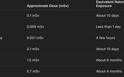
For comparison:
- A CT scan of the abdomen can be around 8–10 mSv.
- A cross-country flight exposes you to about 0.03–0.05 mSv.
Most standard X-rays expose you to only a small fraction of your annual natural background radiation.
Is X-Ray Radiation Dangerous?
In medical imaging, radiation is used in very low doses. For most adults, the risk from a single diagnostic X-ray is extremely small.
The risk depends on:
- The part of the body being imaged
- The number of images taken
- Age (children are more sensitive)
- Frequency of repeated exposure
Healthcare providers follow the ALARA principle (As Low As Reasonably Achievable), meaning radiation doses are minimized while still producing clear diagnostic images.
Are Children at Higher Risk?
Yes, children are more sensitive to radiation because:
- Their cells divide more rapidly.
- They have more years ahead for potential long-term effects to develop.
For this reason, pediatric imaging uses adjusted lower radiation doses, and unnecessary scans are avoided.
What About Pregnant Patients?
Radiation exposure during pregnancy is carefully managed. While a single X-ray (especially of areas away from the abdomen) usually poses very low risk, doctors take extra precautions:
- Shielding the abdomen when possible
- Avoiding non-essential imaging
- Considering alternative imaging like ultrasound or MRI
Always inform your healthcare provider if you are pregnant or might be.
How Does X-Ray Radiation Compare to CT Scans?
It’s important not to confuse X-rays with CT scans.
Imaging Type
Radiation Level
Standard X-ray
Low
CT Scan
Higher (often 10–100 times more than X-ray)
CT scans provide more detailed cross-sectional images but use significantly more radiation. That’s why doctors typically start with X-rays when appropriate.
Does Radiation Build Up Over Time?
Radiation exposure from medical imaging is cumulative over a lifetime. However, occasional diagnostic X-rays contribute only a small amount to overall lifetime exposure.
Medical professionals carefully weigh:
- The benefit of diagnosing a condition
- The minimal radiation risk
In most cases, early detection of fractures, infections, or serious disease far outweighs the tiny radiation risk.
What to Expect During an X-Ray
If you're worried about radiation, knowing what happens during the procedure may help.
Before the Exam
- Remove metal objects and jewelry.
- Inform the technician if you are pregnant.
During the Exam
- The technologist positions you correctly.
- You remain still for a few seconds.
- A brief burst of radiation captures the image.
The exposure itself lasts less than a second.
After the Exam
- There is no radiation left in your body.
- You can resume normal activities immediately.
- Results are reviewed by a radiologist.
How to Minimize Radiation Risk
While medical X-rays are safe, you can take simple precautions:
- Keep a record of previous imaging.
- Ask if alternative tests (like ultrasound or MRI) are appropriate.
- Ensure imaging is medically necessary.
- Choose accredited imaging centers.
Healthcare providers already take significant measures to protect patients, including shielding and modern low-dose equipment.
Final Thoughts
So, how much radiation is in an X-ray? In most cases, the dose is very small — often comparable to a few days or weeks of natural background radiation. Modern imaging equipment uses carefully controlled, minimal radiation to ensure patient safety.
When medically necessary, the benefits of accurate diagnosis far outweigh the tiny radiation risks. If you have concerns, talk to your healthcare provider — they can explain why the test is needed and how safety measures are applied.
Frequently Asked Questions
A chest X-ray typically involves about 0.1 mSv, which is roughly equal to 10 days of natural background radiation.
For most adults, a single X-ray poses extremely low risk.
No. Radiation passes through your body instantly and does not remain afterward.
There is no fixed number. Doctors consider cumulative exposure and medical necessity before ordering repeat imaging.
Yes. Dental X-rays involve very small radiation doses — often less than a day’s worth of natural background exposure.
You should not avoid medically necessary X-rays. Delaying diagnosis may pose greater health risks.
No. Airport body scanners expose you to extremely small radiation levels, typically lower than medical X-rays.