X-Ray of the Hand: Detecting Fractures, Arthritis, and Bone Abnormalities
2/11/2026
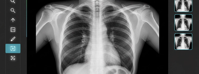
A hand injury or ongoing pain can be both alarming and limiting. When your hand doesn’t feel right, a doctor may order a hand X-ray to look inside. This imaging test is quick, painless, and often the first step in diagnosing a range of bone and joint problems. A hand X-ray lets doctors see the bones, joints, and alignment of your hand in detail. It is one of the most commonly used tools in musculoskeletal care.
In this article, we’ll explain how a hand X-ray works, what it can detect, and why it’s so useful when you have pain, injury, or stiffness in your hand. The goal is to give you a clear, friendly explanation you can understand—even without medical training.
What Is a Hand X-Ray?
An X-ray is a form of imaging that uses a small amount of radiation to take pictures of the inside of your body. When X-rays pass through your hand, dense tissues like bone absorb most of the rays and appear white on the image. Softer tissues like muscles and fat absorb less and look gray or black.
During a hand X-ray, you’ll place your hand on a flat surface. A trained technician will position your hand in different ways so multiple views can be captured. These views help doctors evaluate not just one angle, but the overall structure of the hand.
Why Doctors Use a Hand X-Ray
A hand X-ray is a valuable tool for diagnosing many conditions. Some of the main reasons doctors order this test include:
- Fractures and breaks in the bones
- Joint changes related to arthritis
- Bone abnormalities such as growths or deformities
- Follow-up evaluation after injury or surgery
Let’s look at what these findings can look like on an X-ray and why they matter.
Detecting Fractures and Injuries
One of the most common uses of a hand X-ray is to confirm a fracture. Breaks in bones are usually easy to spot on an X-ray because they show up as clear lines or gaps in the normal bone outline.
Common Hand Fractures
Fractures can occur in many places in the hand, including:
- Metacarpal bones (long bones of the hand) — for example, boxer's fractures near the little finger knuckle are a frequent injury.
- Finger bones (phalanges)
- Carpal bones (small bones at the base of the hand) — fractures like scaphoid breaks are common after a fall with an outstretched hand.
Hand X-rays can also show dislocations, where a bone is pushed out of its normal position. This can be visible as misalignment in the joints.
In some cases, tiny fractures may be hard to see on the first X-ray, especially if bones overlap in the image. If pain and swelling continue despite a normal initial X-ray, your doctor might order further imaging like a CT scan or MRI to get a clearer view.
Spotting Arthritis and Joint Diseases
Hand X-rays are also very helpful in diagnosing arthritis and other joint disorders. Even though cartilage (the cushioning tissue between bones) doesn’t show up directly on X-rays, doctors can infer its condition by looking at the spaces between bones in the joints.
Signs of Arthritis on X-Ray
An X-ray image can show several features that suggest arthritis, including:
- Narrowed joint spaces, which happen when cartilage wears thin
- Bone spurs — extra bony growths that form around joints
- Erosions and irregular bone surfaces seen in conditions like rheumatoid arthritis
For example, in rheumatoid arthritis, the immune system mistakenly attacks the joints, leading to bone erosion and deformity. These changes are often visible on X-ray and help doctors assess severity and track disease progression.
Arthritis can make joints stiff, painful, and difficult to move. By comparing X-ray images over time, doctors can see whether joint damage is getting worse or if treatment is helping.
Detecting Other Bone Abnormalities
Beyond fractures and arthritis, hand X-rays can reveal a range of other bone issues. These include:
- Bone growths or tumors – unusual patterns or shapes may suggest benign cysts or, in rare cases, malignancies
- Congenital abnormalities, such as extra fingers (polydactyly) or fused bones (syndactyly)
- Healing progress, where doctors check how well a broken bone is mending
In children, X-rays are also used to assess bone development and growth plate status. This helps doctors understand whether bones are growing normally.
Limitations of Hand X-Rays
While hand X-rays are extremely useful, they have limits. Most importantly, they do not show soft tissues such as ligaments, tendons, or nerves. For example, a tendon injury can cause pain and swelling without showing up on an X-ray. In such cases, other imaging techniques like MRI or ultrasound may be more informative.
Also, early stages of some conditions—especially certain types of arthritis—may not yet produce visible changes on an X-ray. That’s why doctors combine X-ray findings with your symptoms and physical exam results.
What Happens After Your Hand X-Ray
After your hand X-ray is taken, a specialist called a radiologist reviews the images. They look for fractures, alignment issues, joint changes, and unusual bone patterns. The results are then shared with your healthcare provider, who explains what they mean for your care and treatment.
Based on what the X-ray shows, your doctor may recommend:
- Casting or splinting for fractures
- Physical therapy for joint stiffness
- Medication or lifestyle changes for arthritis
- Further imaging if the X-ray is inconclusive
The process is designed to get you accurate answers and help you move toward recovery.
Conclusion: Why Hand X-Rays Matter
A hand X-ray is a fast and powerful diagnostic tool. It allows doctors to:
- Confirm or rule out fractures
- See signs of arthritis and joint damage
- Detect bone abnormalities and growths
- Track healing over time
While it doesn’t show all tissue types, especially soft tissue injuries, an X-ray gives clear insight into bone health and joint structure. Understanding what an X-ray can reveal helps you take an active role in your care and work with your doctor to find the right path to healing.
Frequently Asked Questions
An X-ray is the primary tool for visualizing the "hard" tissues of the hand. It is most effective at identifying: Fractures: Breaks or cracks in the phalanges (fingers), metacarpals (palm), or carpals (wrist). Arthritis: It shows joint-space narrowing, bone spurs, and erosions, characteristic of Osteoarthritis or Rheumatoid Arthritis. Bone Abnormalities: These can be bone cysts, tumors, or metabolic bone diseases. Foreign Bodies: Detecting metal or glass fragments embedded in the tissue.
Generally, no. X-rays use radiation that passes through soft tissue easily but is absorbed by dense bone. While a doctor might suspect a ligament tear based on how the bones are aligned (or misaligned) on the film, the actual soft tissue will not be visible. For injuries like a "skier’s thumb" or tendon ruptures, an MRI or Ultrasound is usually required.
Preparation is minimal. The most important step is to remove all jewelry, including rings, watches, and bracelets. Metal creates "artifacts" on the image—bright white streaks that can hide a hairline fracture or a small bone abnormality. If a ring is stuck due to swelling, notify the technician; they may have tools to work around it or may need to assist in its removal.
To get a full 3D understanding of your hand's anatomy from 2D images, technicians usually take three standard views: AP (Anteroposterior): Your hand is flat on the plate, palm down. Oblique: Your hand is tilted at a 45-degree angle (often supported by a foam wedge). Lateral: Your hand is placed on its side, usually with the thumb up.
Gemini said Getting an X-ray of the hand is a quick, painless process, but it’s natural to have questions about what the images actually reveal. Whether you've taken a tumble or are dealing with persistent joint pain, here are the most frequently asked questions regarding hand radiography. Hand X-Ray: Frequently Asked Questions 1. What specific conditions can a hand X-ray detect? An X-ray is the primary tool for visualizing the "hard" tissues of the hand. It is most effective at identifying: Fractures: Breaks or cracks in the phalanges (fingers), metacarpals (palm), or carpals (wrist). Arthritis: It shows joint space narrowing, bone spurs, and erosions characteristic of Osteoarthritis or Rheumatoid Arthritis. Bone Abnormalities: This includes bone cysts, tumors, or metabolic bone diseases. Foreign Bodies: Detecting metal or glass fragments embedded in the tissue. 2. Can an X-ray show ligament or tendon injuries? Generally, no. X-rays use radiation that passes through soft tissue easily but is absorbed by dense bone. While a doctor might suspect a ligament tear based on how the bones are aligned (or misaligned) on the film, the actual soft tissue will not be visible. For injuries like a "skier’s thumb" or tendon ruptures, an MRI or Ultrasound is usually required. 3. How should I prepare for the procedure? Preparation is minimal. The most important step is to remove all jewelry, including rings, watches, and bracelets. Metal creates "artifacts" on the image—bright white streaks that can hide a hairline fracture or a small bone abnormality. If a ring is stuck due to swelling, notify the technician; they may have tools to work around it or may need to assist in its removal. 4. What do "AP," "Lateral," and "Oblique" views mean? To get a full 3D understanding of your hand's anatomy from 2D images, technicians usually take three standard views: AP (Anteroposterior): Your hand is flat on the plate, palm down. Oblique: Your hand is tilted at a 45-degree angle (often supported by a foam wedge). Lateral: Your hand is placed on its side, usually with the thumb up. 5. Is the radiation exposure dangerous? The radiation dose for a hand X-ray is extremely low—roughly 0.001 mSv. To put that in perspective, that is about the same amount of natural background radiation you receive from the environment in just three hours of daily life. While you should always inform your doctor if you are pregnant, a hand X-ray is considered one of the safest imaging procedures available.