Knee X-Ray: Anatomy, Procedure & What to Expect
2/11/2026
The knee is a vital joint that lets us walk, run, climb stairs, and carry out everyday movements. When you experience pain, swelling, or trouble bending your knee, a knee X-ray is often one of the first tests doctors use to find the cause. It’s fast, simple, and usually painless. This article will explain what a knee X-ray shows, why it’s ordered, how it works, and what you can expect from start to finish.
What Is a Knee X-Ray?
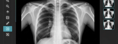
A knee X-ray is a diagnostic imaging test that uses a small amount of radiation to create detailed pictures of the internal structures in and around your knee joint. The images are black and white: dense structures like bones show up in white, while softer tissues appear as shades of gray.
This test focuses on the major bones of the knee, including:
- Femur (thigh bone)
- Tibia (shin bone)
- Fibula (smaller lower leg bone)
- Patella (kneecap)
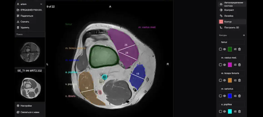
While X-rays capture bone clearly, they do not show soft tissues like ligaments, tendons, or cartilage in detail. For those, an MRI or ultrasound might be needed later.
Why You Might Need a Knee X-Ray
Doctors commonly recommend a knee X-ray in the following situations:
1. To Investigate Pain or Injury
If you’ve had trauma to the knee — like a fall, twist, or blow — an X-ray can show fractures, dislocations, or bone injuries. It helps doctors diagnose the problem quickly.
2. To Assess Joint Alignment and Structure
When your knee feels unstable or you notice a visible abnormality, an X-ray helps check for irregular joint positions or bone deformities.
3. To Detect Arthritis or Wear-and-Tear
X-rays can reveal changes in the joint space, bone spurs, and signs of osteoarthritis — wear and tear that causes pain and stiffness over time.
4. For Follow-Up After Treatment
If you’ve had surgery or a fracture has been set, follow-up X-rays can show how well the bone is healing and whether implants are in the correct position.
5. To Rule Out Bone Conditions
In some cases, X-rays help detect infections, cysts, or tumors in the bone.
How to Prepare for Your Knee X-Ray
One of the benefits of a knee X-ray is that it requires very little preparation:
- Wear comfortable clothing that doesn’t contain metal, such as zippers, buttons, or snaps — metal can show up in the images and affect clarity.
- You may be asked to remove jewelry around the knee or lower body.
- If you are pregnant or think you might be, tell your healthcare provider before the test. While the radiation dose is low, your doctor will make decisions to protect your baby.
There’s no need to fast, stop medications, or take special steps before a knee X-ray.
What Happens During the Procedure
Getting a knee X-ray only takes about 10–15 minutes from start to finish.
Here’s what typically happens:
- Positioning: You will either stand, sit, or lie down, depending on the views needed.
- Multiple Views: The technician will take several images from different angles — usually a front view (AP), a side view (lateral), and often a kneecap view (sunrise) to get the most complete picture of the joint.
- Stillness: You’ll be asked to stay still and sometimes to hold your breath briefly while each image is taken. Movement can blur images, so staying still helps ensure clear results.
- Non-Painful: You won’t feel anything from the X-ray. However, if your knee hurts, holding certain positions might be uncomfortable. Inform the technologist right away if any position causes pain.
After the X-Ray: Results and Follow-Up
Once your images are captured, the technologist checks them to make sure they are clear and complete. A radiologist — a doctor trained in interpreting imaging — reviews the X-rays and sends a report to your referring healthcare provider.
Depending on your facility, results may be available within minutes (especially in urgent settings) or a couple of days. Your doctor will go over the findings with you and discuss your treatment options. Treatment might range from rest and physical therapy to medication, braces, or additional imaging if a soft-tissue issue is suspected.
Safety and Risks
A knee X-ray involves a very low dose of radiation, much less than many other imaging tests. For most people, the benefits of accurate diagnosis greatly outweigh any small risk from radiation exposure.
Still, if you are pregnant or have concerns about radiation, let your healthcare provider know. They may use protective shields or adjust the procedure as needed.
Limitations of Knee X-Rays
While X-rays show bones and joint spaces clearly, they have limitations:
- Soft tissues like ligaments, tendons, and cartilage are not visible in detail on standard X-rays.
- Early cartilage damage or meniscal tears may not appear, so your doctor might order an MRI if these are suspected.
Despite these limitations, X-rays remain a fast, inexpensive, and widely available first step in diagnosing knee problems.
Frequently Asked Questions
A knee X-ray is very quick - typically around 10–15 minutes from positioning to completion.
No, the X-ray procedure itself is painless. Pain might come from holding a certain position if your knee is already sore.
X-rays show bones, joint spacing, fractures, dislocations, and signs of arthritis. They cannot show soft tissue tears like ligament injuries.
No special preparation or fasting is needed. Just wear comfortable clothing without metal over your knees.
The radiation dose from a knee X-ray is very low. For most people, it’s considered safe when medically necessary. Tell your provider if you are pregnant to ensure appropriate precautions.